IN - SITU NEUTRON REFLECTIVITY STUDY
OF SURFACE – INDUCED ORDERING OF MAGNETIC COLLOIDS
A. Vorobyov 1 , J. Major 1 , H. Dosch 1
, G. Gordeev 2
, D. Orlova 2 .
1.
MPI-MF Stuttgart, Germany.
2.
PNPI St - Petersburg, Russia.
Understanding the internal structure of the magnetic colloids (MC) is an attractive topic not only because of the increased interest in objects of nanoscopic scale. It is also a part of a fundamental question regarding the modification of the structure of simple and complex liquids in constrained geometry’s. Despite of a number of studies on MC, the micro-structure of a MC in the vicinity of its surface is an open problem. In particular, is the interface inducing an unusual particle ordering changing tribological or magnetic properties of MC? It has been recently shown [1] that specular neutron reflection is one of the most effective methods to study MC microstructure in a layer close to a buried interface, in view of the strong penetrating ability of neutron radiation and its sensitivity to nano-scale inhomogeneities.
We report on neutron specular reflection experiments performed on a D2O-based MC wetting single-crystalline Si substrate. The samples were fabricated at Petersburg Nuclear Physics Institute (PNPI). Single domain ferromagnetic grains made by chemical deposition of dispersed magnetite (Fe3O4) were coated with a surfactant (sodium oleat C18H33O2Na) and dissolved in heavy water with a magnetite volume concentration C @ 6 %. We found by small angle neutron scattering, that the mean diameter of the coated grains is of the order dm = 100 Å. In analysis of this SANS data it was assumed that the scatters have a spherical shape, this was confirmed by atomic-force microscopy measurements that however showed a significant (about 50 %) polydispersity of the samples.
Neutron reflectivity experiments were carried out at the EVA reflectometer-diffractiometer [2] at the Institute Laue-Langevin (ILL, Grenoble, France). The sample holder was an open quarts frame glued on the Si single crystal block. Neutron reflectivity measurements on the bare substitute indicated that the polished surface was covered by a SiO2 layer (15 – 25 ) Å thick. The reflecting plane of EVA is vertical, a geometry which allows the determination of the reflectivity from the bottom Si – MC interface. The primary beam reaches the interface through the side of the Si head as shown in Figure 1a. The reflected, transmitted, and scattered intensities are simultaneously recorded by a linear position-sensitive detector. A homogeneous magnetic field up to 110 Oe was applied parallel (H║) or perpendicular (H┴) to the interface by means of a pair of Helmholtz coils. The data showed evidence of ordering in the proximity Si. A strong Bragg diffraction peak (Figure 2b) denotes that in the volume close to the SiO2 surface MC particles built a stable multilayer structure similar to that of smectic liquid crystals. This layering process depends on the sample concentration, direction and strength of the external magnetic field, the temperature of the sample.
When the magnetic field is applied parallel to the interface, the Bragg peak is always located at a momentum transfer value corresponding to the interlayer distance L = 100 Å, which is equal to dm. Increasing the magnetic field results only in an increasing of the Bragg peak amplitude. This means that H║ broadens the region of coherent layering. The process is reversible and can be repeated a few times on the same sample.
Quite a different layering configuration is obtained when the magnetic field is applied perpendicular to the interface. At H┴ = 52 Oe, L = 64 Å, while at H┴ = 105 Oe, L = 55 Å, i. e. interlayer distance is about half of dm (see Figure 1 b, c). The size of the peak suggests that the layers are compressed rather than interdigitated, and the dynamics of the process is such that equilibrium is achieved after several hours.
Neutron reflectivity measurements have dramatically shown how magnetic fields alter the structural ordering of MC in constrained geometry. Proceeding in a systematic way from these sparse observations, we are confident that a detailed picture can emerge of the kinetics of such nanoscopic ensembles.
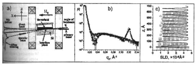
Figure 1.
a)
Sketch of the
experiment; b) reflectivity curves from the interface Si-FF6
in zero field (○) and in H┴ = 105 Oe (●); c) Solid and dash line are
appropriate fit results.
References:
1. A. Vorobyov, et al. J. Appl. Phys. A 74 Suppl. 1 (2002) 81; A. Vorobyov, et al. Physica B 297 (2001) 194; B. P. Toperverg, et al. Physica B 283 (2000) 203.
2. H. Dosch, et al. Rev. Sci. Instrum. 63 (1992) 5533; http: //www.ill.fr/YellowBook/EVA/