APPLICATION OF
MAGNETIC DEVICES AND
SORBENTS
IN GASRTOINTENSTINAL SURGERY
Kanshin
N. N.1, Chikov V. M.2, Cherkasova O. G.3,
Tsybusov S. N.4, Kochenov V.
I.4
1. N. V. Sklifosovskiy Scientific Research Institute of Emergency Medicine, Russian Federation, Moscow, Sukharevskaya place, .
2. N. M. Emanuel Institute of biochemical physics RAS, Russian Federation, Moscow, 119991, Kosygina street, 4.
3. I. M. Sechenov Moscow Medical Academy, Russian Federation, Moscow, Pirogovskaya street,.
4. Medical State Academy, Russian Federation, Nizhniy Novgorod, 603104, Medicinskaya street, 1.
Introduction.
We have been working on application of magnetomechanical forces in abdominal surgery since 197 [1 – 6] and continue our work in this area.
Significant
difficulties in gastrointestinal surgery are caused by non-sterility of the
inner surfaces of gastrointestinal tract organs and their contents. The common
task during such surgery is formation of anastomoses: joining incised hollow
organs while preserving connectivity of their internal cavities. Such
connections should be hermetic and the joined organs should touch each other
only with their sterile serous (outer) surfaces. The situation is complicated
even further by the relatively thin and mechanically weak walls of many of the
organs. Imperfect connections lead to numerous complications: inflammations,
nonhermeticity, impossibility and peritonitis. Several technologies of seamless
connections of holloworgans have been developed over the last decades to
overcome these difficulties, including different glues and various mechanical
devices, which connect the tissue using elastic forces, forming so-called
compressing anastomoses. Each of these methods has advantages and
disadvantages, but the problem of flimsiness of such joints remains to be
solved because of difficulties in providing hermetically, sterility of joints
and matching of serous membranes.
Materials and methods.
We solve these problems using magnetic devices based on permanent magnets. The principle scheme of the operation is illustrated in Figure 1. The typically ring-shaped devices are inserted inside the hollow organs being connected. The magnetic elements are mutually attracted to each other, squeezing tissues between them. A hermetic seal is formed, and the possibility of the gastrointestinal tract is maintained. The patient can eat immediately after operation.
Tissues
squeezed between the elements necroses in 7 – 10 days and inoculation of serous
membranes of the joint organs occurs along the outer (external) perimeter of
the elements. After necrosis of the squeezed tissues the pressing elements are
separated and excreted from the organism through the natural way.
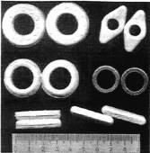
We designed and tested many types of pressing magnetic elements (Figure 2): rigid and elastic, of different sizes and shapes (rings, prisms, rhombi), with special arrangements for tissue fixation, etc. Typically multi-polar magnetic systems were used, since they can be made elastic and provide sufficient force with minimal size and weight. We used SmCo and NdFeB permanent magnets, imbedded in a silicone plastic to provide biocompatibility and elasticity. Surgical techniques of the magnetic device applications for different types of operations were also developed.
52 mongrel dogs were used in pre-clinical trials. Two to four anastomoses were performed on each of the animals. Kinetics of tissue compression between the magnetic elements and the following necrosis, as well as tissue regeneration and joint formation, was studied. Histological studies were also performed. Clinical trials of the magnetic devices were performed on more than 100 patients at the N. V. Sclifosovskiy Moscow Scientific Research Institute of Emergency Medicine.
|
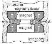
Figure
1. Scheme of joining hollow organs using ring-shaped magnetic devices. A
indicates the zone of tissue growth and anastomosis formation. White arrows
indicate magnetization direction. |
Figure
2. Magnetic devices used for forming anastomoses of hollow organs of
abdominal cavity. Scale is in centimeters. |
Results and discussion.
Both pre-clinical and clinical data indicate good quality of the anastomoses formed by the magnetic devices and the ease of use of the later.
The animal experiments have shown, that necrosis and rejection of the tissues squeezed by the magnets is not a mechanical amputation, but a complex biophysical process. The pressed tissue loses blood supply, and the oxygen-starved cells are partially destroyed by the pressure and die of both hypoxia and mechanical damage. The products of cell destruction moving from the zone of pressure stimulate the active rejection of the dead tissues by the organism (including immune reactions). The more intensive is the pressing force, the faster the rejection. It was found, that tissue regeneration occurs mostly in the zone of moderate pressure at the periphery of the magnetic devices. A mathematical model of the first stages of the pressing process was suggested. Its predictions correlated well with experimental data.
Detailed morphological and histological investigations demonstrated the abscence of any harmful influence of the magnetic field on the joined tissue and organs.
The presence
of high-gradient magnetic field at the site of operation allows the
organization of a targeted drug delivery, either through the bloodstream (this
could be beneficial for antibiotics, for example) or through the
gastrointestinal tract. We conducted successful experiments of targeted delivery
of catalyses (for faster dead tissue destruction) conjugated with ferro-carbon
particles through the gastrointestinal tract.
Clinical trials of the magnetic devices were successfully performed at the N. V. Sclifosovskiy Moscow Scientific Research Institute of Emergency Medicine during operations on various parts of the human gastrointestinal tract, including gullet, esophagus, Stomach, duodenum, bile ducts, large and small intestine, and rectum. Magnetic elements were also successfully applied in urology. Overall more than 100 patients were treated. All operations were successful, with low occurrence of mild after-operation complications. A high quality of anastomoses was observed: an absence of scars or narrow spots and a low occurrence of inflammations. After separation of the magnetic devices no artificial objects (threads, stiches, etc.) remained in the patients’ organism. Surgeons easily learned the technique and found it to be convenient. Application of the magnetic devices reduced the duration of the operation and lowered the requirements for the surgeon qualification. All these factors increase the reliability of the operation and reduce the risk of complications.
References.
1. Kanshin N. N., Tsybusov S. N., Cherkasova O. G., Kochenov V. I., Chikov V. M. Method of hollow organs tissue connection. // Patent of USSR № 2111068, 1971.
2. Kanshin N. N., Gleizer V. M., Hutoryanskiy A. A., Chikov V. M. Device for tissue connection. // Patent of USSR № 8735063, 1975.
3. Kanshin N. N., Permyakov N. K., Chikov V. M. Studies of the tissue compression process (in Russian). // Archive of Pathology, 8 (1975), 378 – 42.
4. Kanshin N. N., Dgalagonia R. A., Chikov V. M. Method of hollow organs tissue connection. // Patent of USSR № 2505398, 1977.
5. Kanshin N. N., Dgalagonia R. A., Chikov V. M. On the tendencies of the processes of tissue compression and regeneration during the formation of «Seamless» anastomoses of hollow organs of the gastro-intestinal tract (in Russian). // Izvestia AN USSR, Ser. Biology, 1 (1979), 13 – 17.
6. Kanshin N. N., Tsybusov S. N., Cherkasova O. G., Kochenov V. I., Lipatov V. A., Hamidov A. I., Gus’kov I. A. Chikov V. M. Device for forming anastomoses of hollow organs. // Patent of USSR, № 1142926, 1983.